ChroMS Multicolor Imaging
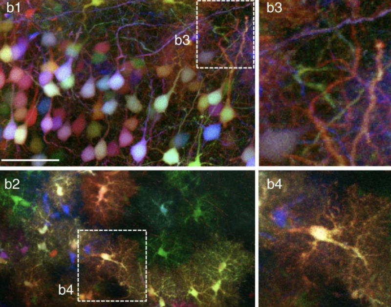
Published today in Nature Comms is a study by Abdeladim and colleagues entitled “Multicolor multi-scale brain imaging with chromatic multiphoton serial microscopy”. Using a hacked TissueCyte, the authors acquire data from four PMT channels whilst exciting using two laser lines. With the pulses of the two lines synchronized, a third virtual line is created. For example, with λ1 = 850 nm and λ2 = 1100 nm a virtual line at 950 nm is created. The “intensity” of this virtual line can be manipulated by slightly altering the phase of the two pulse trains. With carefully chosen excitation optics, chromatic aberration is minimized to allow high resolution imaging across a wide field of view. The approach is show-cased using beautiful Brainbow-labeled tissue. Spectral unmixing allows about 20 different colors to be distinguished.